
Asbestos, once lauded for its fire-resistant properties, has left a pernicious legacy affecting countless individuals across the globe. The insidious nature of asbestos exposure has prompted a crucial demand for reliable diagnostic measures to detect its impact on human health. This essay will illuminate the science behind biomarkers of asbestos exposure, which serve as critical indicators within biological specimens, signaling the presence of this hazardous material. We will explore how specific proteins and DNA alterations within blood, urine, and lung tissue offer insights that not only help diagnose asbestos-related conditions but also chart the history and severity of exposure. Additionally, the essay will examine how radiological and pulmonary function tests provide a lens through which the medical community can visualize and assess the resultant lung damage, shaping treatment strategies and surveillance efforts for those affected by this harmful mineral.
Biomarkers of Asbestos Exposure
Identifying Biomarkers of Asbestos Exposure
In the realm of occupational health and pulmonary medicine, the detection and analysis of asbestos exposure biomarkers represent a critical pursuit. Asbestos, a group of naturally occurring fibrous minerals, has been ubiquitously used for its durability and resistance to heat, but its fibers can become deadly when inhaled, leading to severe health complications such as asbestosis, lung cancer, and mesothelioma. Detecting asbestos exposure in individuals has substantial implications for early intervention and disease prevention.
Biomarkers that are indicative of asbestos exposure have become a focal point for early detection strategies. Soluble mesothelin-related peptides (SMRPs) and osteopontin represent two such serum biomarkers. SMRPs are elevated in individuals with mesothelioma, an aggressive form of cancer associated with asbestos exposure. Osteopontin, a glycoprotein found in bone and other tissues, has also been associated with asbestos exposure and mesothelioma, although its specificity is relatively lower. The quantification of these biomarkers, alongside high-resolution radiographic imagery, augments the clinical assessment of asbestos-induced conditions.
Another approach to ascertaining asbestos exposure involves examining the prevalence of asbestos bodies in bronchoalveolar lavage fluid or lung tissue biopsies. Asbestos bodies are asbestos fibers coated with an iron-containing proteinaceous material, a response by the human body to the inhaled fibers. When found in significant numbers, asbestos bodies serve as a definitive indicator of asbestos exposure, albeit not without ambiguity regarding the timing and dose. Furthermore, recent studies have been investigating the role of microRNAs, small non-coding RNA molecules that regulate gene expression, as potential biomarkers, paving the way for innovative diagnostic modalities. The aggregate of these biomarkers contributes to a comprehensive understanding of asbestos exposure and its potential health ramifications.

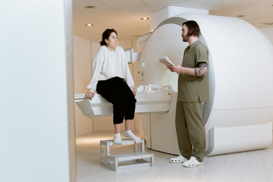
Radiological and Pulmonary Function Testing
Radiological assessments, such as chest X-rays and computed tomography (CT) scans, are pivotal in the visual evaluation of the lung parenchyma following suspected asbestos exposure. Specifically, CT scans offer superior clarity and detail, enabling the detection of small, early-stage pleural and parenchymal abnormalities often invisible on standard radiographs. These may include pleural plaques, thickening, and calcifications—hallmarks of asbestos-related disease. Moreover, both chest X-rays and CT scans are efficacious in identifying fibrotic changes indicative of asbestosis, a chronic lung disease resulting from asbestos fiber inhalation.
While radiological imaging does not quantify exposure or predict disease development with precision, it serves as a tool for diagnosing manifestations of asbestos-related lung pathology, including malignant mesothelioma and lung cancer.
Pulmonary function tests (PFTs) complement radiological methods by providing quantifiable data on the impact of asbestos fibers on respiratory mechanics. These tests measure lung volumes, capacities, rates of airflow, and gas exchange, which are often impaired in asbestotic processes. Restrictive lung defects characterized by reduced lung volumes, particularly forced vital capacity (FVC), are a common finding. PFTs may also reveal a reduced diffusing capacity for carbon monoxide (DLCO), reflecting impaired gas transfer across the alveolo-capillary membrane—a consequence of fibrotic tissue replacement. Although pulmonary function tests cannot conclusively diagnose asbestos-related disease or discern past exposure levels, they are crucial for assessing functional impairment and monitoring disease progression or response to therapy in affected individuals.
Recognizing the subtle cues left behind by asbestos exposure is a complex yet vital process in protecting public health. The biomarkers and diagnostic tools discussed underscore the scientific community’s capability to not only identify those who have been exposed but also monitor their health conditions with precision. As we deploy these advanced methodologies, from analyzing biomarkers to harnessing the power of imaging and pulmonary tests, we equip ourselves with the knowledge needed to mitigate the consequences of exposure. These strategies, rooted in robust research and clinical inquiry, shine a beacon of hope onto a path riddled with the challenges of past environmental missteps, guiding us toward a future where the insidious threats of asbestos are significantly diminished.